
Microscope Slides
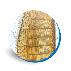
Corn Root Tip
Corn also known as “Maize” is a monocot and has a fibrous root system. The roots of a maize plant spread to about 4.9ft laterally and up to 6.5ft downwards.
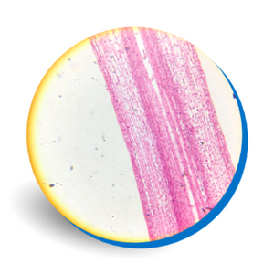
The fibrous root types consume nutrients and dampness near the soil surface to provide a broader footprint under the corn plant that helps maximize nutrient and water uptake. Moreover, corn roots have deep roots – that help find any trace of water within reach.
Observe and explore this prepared slide of corn root cap followed by the zone of cell division, zone of elongation and zone of maturation.
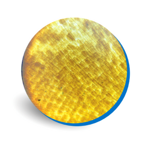
Pumpkin Stem
This is widely known as the pumpkin’s stem or handle, but the technical term is “peduncle”. It links the pumpkin to the vine. The quality of the pumpkin can be determined through the quality of the peduncle.
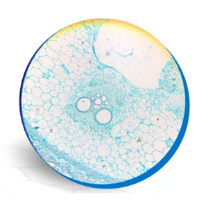
The stem is green through the growing cycle and as the fruit ripens, it slightly curved and turns to brownish green. The peduncle is the umbilical cord bringing nutrients to grow the pumpkin fruit.
It’s made of the xylem – specialized tissue composed of protoxylem and metaxylem. Pumpkin stems are atypical for dicots since they have phloem on both sides of the vascular bundle.
Notice the large vascular bundles and thick epidermis of the pumpkin in this dicot stem microscope slide.
Pine Leaf
This slide shows the cross section of pine leaf. Under the microscope, pine leafs reveal a complex structure that is vital to the pine tree.
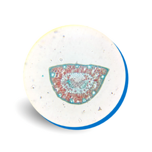
The fibrovascular bundles, serves like blood vessels in the human body. Part of this bundle is the xylem which transfers water and minerals to the plant, and the other is called phloem which moves the nutrients downward from the leaves.
Resin ducts, hold the resin of the pine tree as a protection against insects and animals. Chlorophyll cells called chlorenchyma, serves as protection of the pine tree from rain and strong wind. Stoma allow the tree to breathe. These are special pores that allow gases to exchange within the cell.
Onion Epidermis
Onions are made up of several layers with a membrane separating each layer. The transparent layers of onion bulb skin does not contain chloroplast and chlorophyll and exists in a single layer of epidermal cells that carry out the function of storing energy.
See and discover onion epidermal cells with this slide. Each plant cell has a cell wall, cell membrane, cytoplasm, nucleus, and a large vacuole. The nucleus is present at the periphery of the cytoplasm. The vacuole is prominent and present at the center of the cell, surrounded by cytoplasm.
Bee Leg
Though small in size, honey bees are complex creatures. Honey bees are very flexible with three pairs of legs split into six segments. The front legs are specially designed to clean the antennae, while the rear legs have a section devoted to pollen accumulation called a pollen basket.
Each leg has claws for gripping and sticky pads to assist the bee in landing on slick surfaces. Bees also have taste receptors on the tips of their legs.
Worker bees have unique and different set of back legs containing an auricle, special combs and a pollen press. These are used to brush, collect, pack and carry pollen and propolis back to the hive.
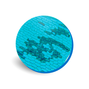
Butterfly Wing
Butterfly wings come in many different shapes, colors, textures, and patterns. Butterfly wings are made of two chitonous layers (membranes) that are nourished and supported by tubular veins.
The veins also function in oxygen exchange. Covering the wings are thousands of colorful scales, together with many hairs (setae).
Lepidoptera in Greek means scale wing. These wing scales are tiny overlapping pieces of chitin on a butterfly or moth wing. The scales are outgrowths of the body wall and are modified, plate-like setae (hairs). The front and back of the wings usually have different patterns.
This slide shows the small, individual scales that make up the patterns we see on butterfly wings. It shows the scales that form the elaborate color patterns.
Mosquito Mouthpieces
A microscope slide showing a Whole Mount of the head of a female mosquito displaying the mouthparts.
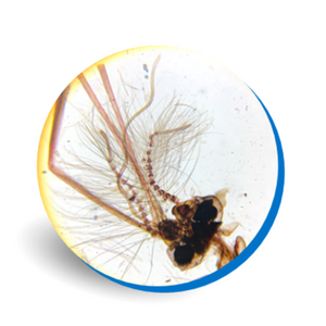
The mosquito's mouth, also called a proboscis is a sophisticated system of six thin, needle-like mouthparts called stylets. The mouthparts of female mosquito are piercing and sucking type. Each of which pierces the skin, finds blood vessels and makes it easy for mosquitoes to suck blood.
The mouthparts include labium, labrum-epipharynx, hypopharynx, mandibles and first maxillae. Only maxillary stylets and mandibular stylets are present in bugs, whereas labrum-epipharynx, hypopharynx, maxillary stylets and mandibular stylets are all present in mosquitos.
Ant
Investigate the anatomy of the ant including head, antennae, eyes and mouthparts.
Ants are social in habit and live together in organized colonies. They range in size from about 0.08 to 1 inch. Their colour is usually yellow, brown, red, or black.
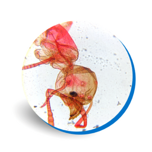
Ant has a large head and a slender oval abdomen joined to the thorax, or midsection, by a small waist. In all ants, there are either one or two finlike extensions running across the thin waist region. The antennae are always elbowed. There are two sets of jaws - the inner pair is used for chewing and the outer pair is used for carrying objects such as food and for digging.
Ants don’t have lungs. Oxygen enters through tiny holes all over the body and carbon dioxide leaves through the same holes.
Frog Blood
Frog's blood is similar in some ways to human's, but different in others.
The red blood cells of frogs are larger than human red blood cells. They are also somewhat elliptical rather than round like human red blood cells.
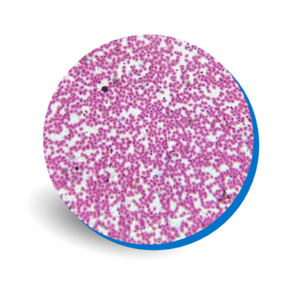
Unlike typical mammalian red blood cells, those from amphibians, such as frogs, contain a DNA-bearing nucleus that is visible in the center of the cell. These cells do divide unlike ours. Frog's white blood cells are very much the same as ours and serve the same function. Frog blood does not have platelets.
Using this slide, observe the frog blood cells under the microscope and see the difference.
Small Intestine
The small intestine is where much of the digestion and absorption of food takes place. It’s the part of the gastrointestinal tract following the stomach surrounded by the large intestine.
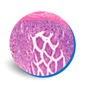
Observe the epithelia lining and villi of the small intestine with this microscope slide. The inner lining of the colon and small intestine is a simple columnar epithelium constantly renewed by the proliferation of stem cells residing within pockets, or crypts, along the intestinal wall.
The villi contain large numbers of capillaries that take the amino acids and glucose produced by digestion to the hepatic portal vein and the liver. Lacteals are the small lymph vessels that are present in villi. They absorb fatty acids and glycerol, the products of fat digestion, into direct circulation.